Emergent Technologies

VIBRANT

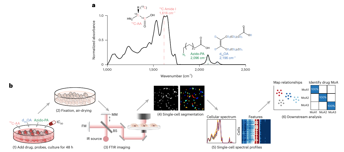
VIBRANT workflow combining vibrational probes, FTIR imaging, single-cell segmentation and downstream drug-response analysis

Drug responses can vary substantially from cell to cell, even within the same tissue or tumor. Population-averaged measurements can therefore provide an incomplete picture of drug efficacy, making single-cell analysis desirable for drug-response testing. However, a practical single-cell platform must combine strong sensitivity with high throughput and rich biochemical information, while remaining affordable, straightforward to operate and robust against batch effects.

VIBRANT, or vibrational painting, addresses these requirements by integrating three infrared-active metabolic probes, mid-infrared imaging and an optimized analysis pipeline. The probes report protein synthesis, saturated fatty-acid metabolism, and unsaturated fatty-acid metabolism, while automated segmentation and machine learning convert each cell's spectrum into a multidimensional drug-response profile. Across more than 20,000 cells and 23 drug treatments, VIBRANT sensitively distinguished mechanisms of action with minimal batch effects, identified compounds with potentially new mechanisms and evaluated drug combinations. This combination of content, scale and accessibility establishes a versatile platform for phenotypic screening and drug discovery.

  • X. Liu, L. Shi, Z. Zhao, J. Shu and W. Min. “VIBRANT: spectral profiling for single-cell drug responses,” Nature Methods 21, 501–511 (2024).
PLANCK

Blablabla blablabla blablabla blablabla blablabla blablabla. Blablabla blablabla blablabla blablabla blablabla blablabla. Blablabla blablabla blablabla blablabla.

Optical Barcoding

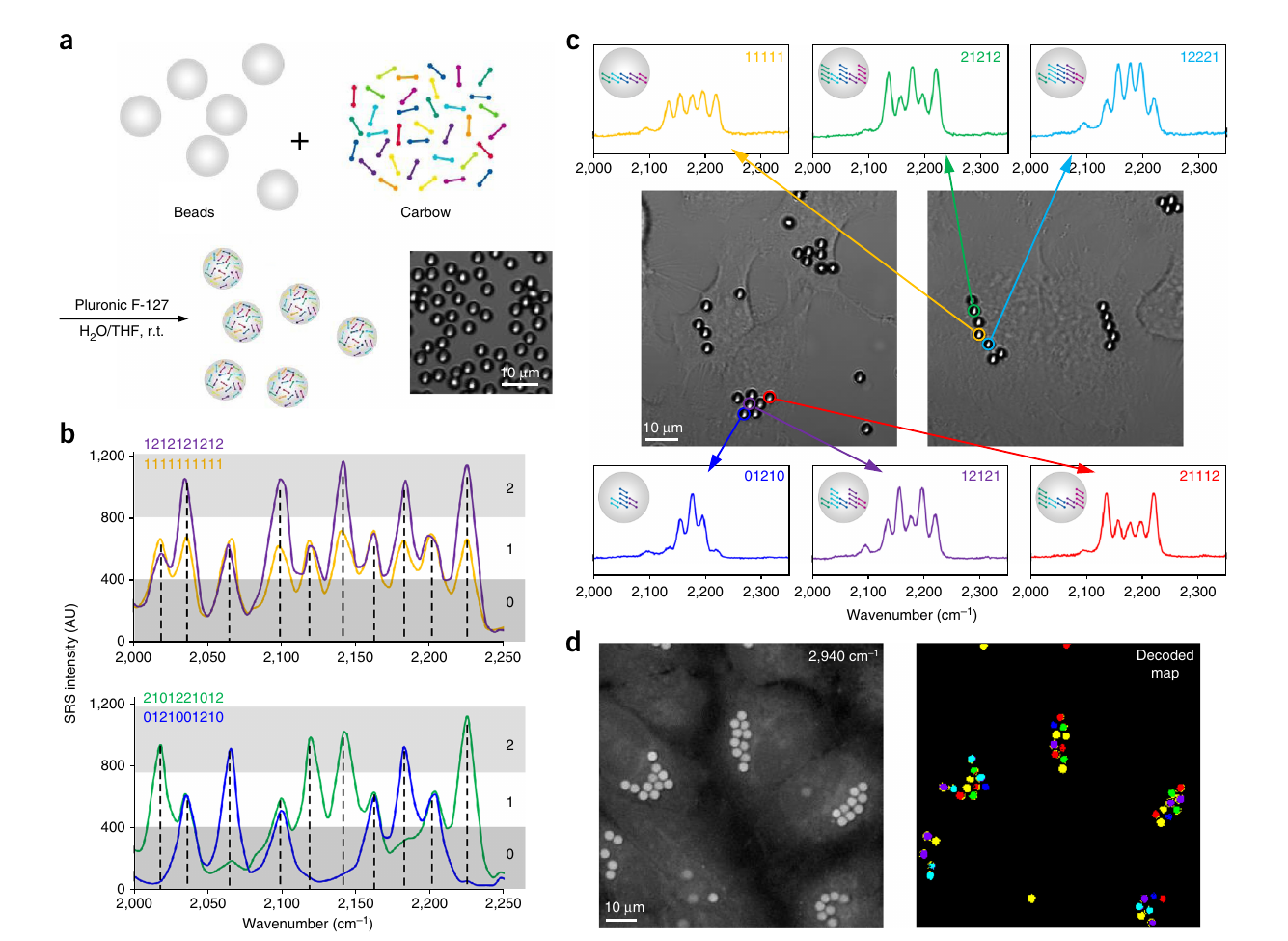
Carbow probes encoding and decoding spectral barcodes in polymer beads and living cells

Systems biology and high-throughput diagnostics increasingly require distinct optical identities for large numbers of cells, biomolecules, antigens and drugs. Conventional fluorescent materials are constrained by broad, overlapping spectra and cross-talk, limiting the number of codes that can be resolved. The Carbow probes in our bioorthogonal imaging toolkit are particularly handy here, providing 20 narrow and independently tunable frequencies that overcome this spectral ceiling.

We encoded microscopic polymer beads using ten Carbow frequencies at three intensity levels. This ternary scheme generates 310-1, or 59,048, distinct spectral barcodes, far beyond the previous record of roughly 1,000. The micron-scale beads can be taken up in multiple combinations by individual cells, where their identities remain readable by spontaneous Raman spectroscopy or can be rapidly mapped by hyperspectral SRS imaging. Combining three encoded beads provides approximately 3 × 10¹³ possible identifiers, sufficient in principle to distinguish every cell in the human body and opening new possibilities for cell tracking, interaction mapping, screening and molecular diagnostics.

  • F. Hu, C. Zeng, R. Long, Y. Miao, L. Wei, Q. Xu and W. Min. “Supermultiplexed optical imaging and barcoding with engineered polyynes,” Nature Methods 15, 194–200 (2018).