Imaging Probes
To realize the full potential of these technological advances, we established a bioorthogonal imaging platform and built a versatile toolkit of compact vibrational probes. These tools bridge the gap between label-free contrast and precise, multiplexed visualization of the molecules that drive living systems.
Bioorthogonal Chemical Imaging
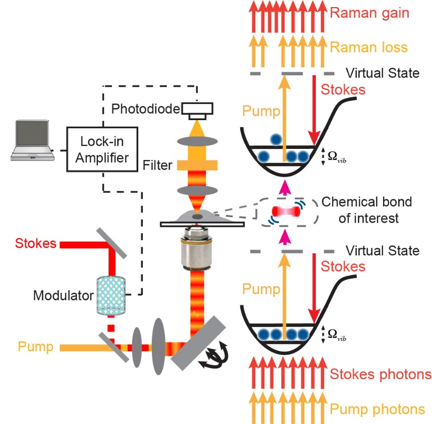
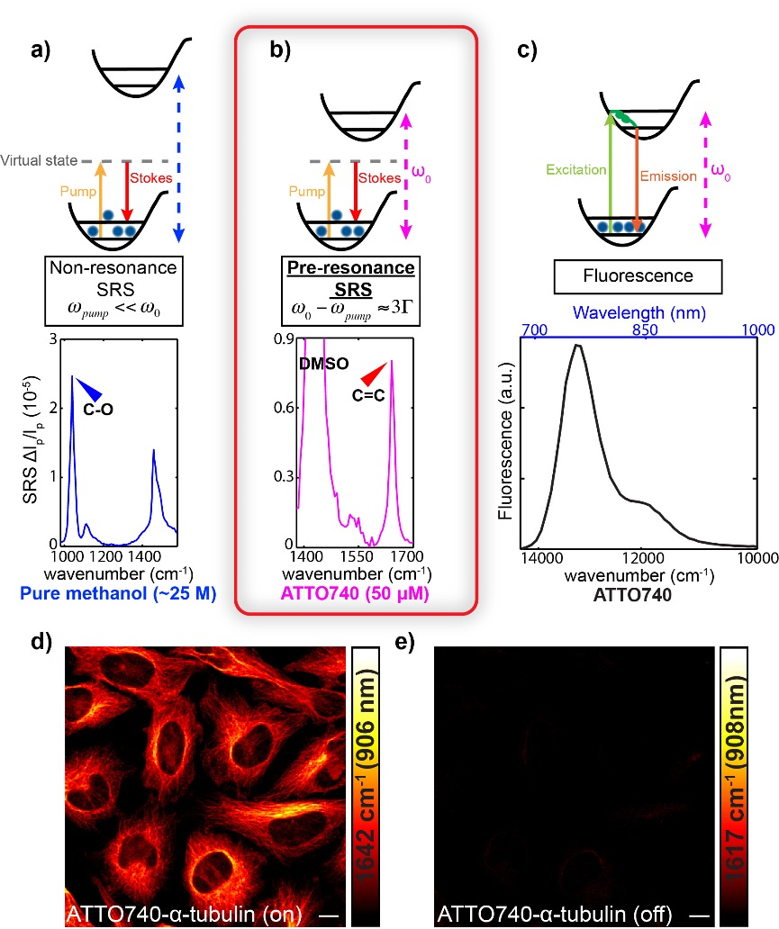
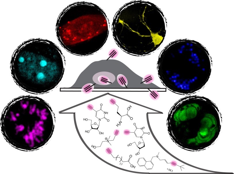
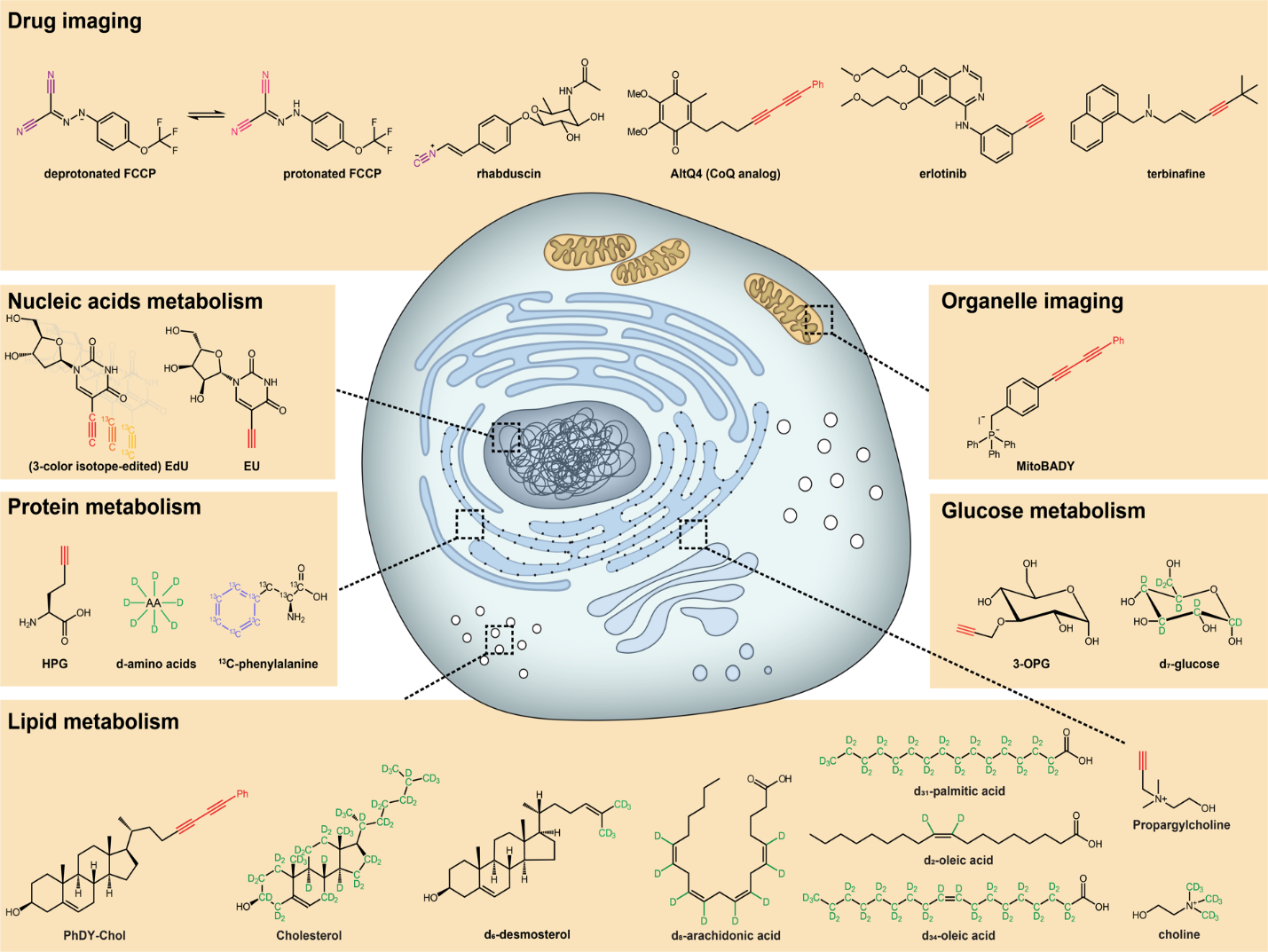
Fluorescence microscopy provides exceptional sensitivity, but its comparatively bulky labels can perturb or obscure the native behavior of small biomolecules. Bioorthogonal chemical imaging addresses this limitation by coupling SRS microscopy with tiny Raman-active tags, including alkynes and stable isotopes, that integrate into biological molecules with minimal disruption.
This platform enables live imaging of nucleic-acid building blocks, amino acids, fatty acids, choline, glucose, cholesterol, and small-molecule drugs across cells, tissues, and model organisms. Its combination of specificity, biocompatibility, and real-time observation opens previously inaccessible views of small-molecule metabolism and dynamics, while multicolor probe strategies extend the approach toward systems-level measurements.
- L. Wei, F. Hu, Z. Chen, Y. Shen, L. Zhang and W. Min. “Live-cell bioorthogonal chemical imaging: stimulated Raman scattering microscopy of vibrational tags,” Acc. Chem. Res. 49, 1494 (2016).
- L. Wei, F. Hu, Y. Shen, Z. Chen, Y. Yu, C. Lin, M. Wang and W. Min. “Live-cell imaging with alkyne-tagged small biomolecules by stimulated Raman scattering,” Nature Methods 11, 410 (2014).
- Z. Chen, V. C. Cornish* and W. Min*. “Chemical tags: inspiration for advanced imaging techniques,” Curr. Opin. Chem. Biol. 17, 637 (2013).
Vibrational Probes for Small Biomolecules
Compact Raman-active tags create distinct spectral signatures without overwhelming the structure or function of their targets. Tunable alkynes, isotopes, and polymer nanoparticles support multicolor live-cell imaging and expand the range of small molecules that can be followed directly.
- Z. Zhao, Y. Shen, F. Hu and W. Min. “Applications of vibrational tags in biological imaging by Raman microscopy,” Analyst 142, 4018 (2017).
- F. Hu, S. D. Brucks, T. Lambert, L. Campos and W. Min. “Stimulated Raman scattering of polymer nanoparticles for multiplexed live-cell imaging,” Chem. Commun. 53, 6187 (2017).
- Z. Chen, D. Paley, L. Wei, A. Weisman, R. Friesner, C. Nuckolls* and W. Min*. “Multicolor live-cell chemical imaging by isotopically edited alkyne vibrational palette,” J. Am. Chem. Soc. 136, 8027 (2014).
- L. Wei, F. Hu, Y. Shen, Z. Chen, Y. Yu, C. Lin, M. Wang and W. Min. “Live-cell imaging with alkyne-tagged small biomolecules by stimulated Raman scattering,” Nature Methods 11, 410 (2014).
Super-Multiplexed Polyyne Probes
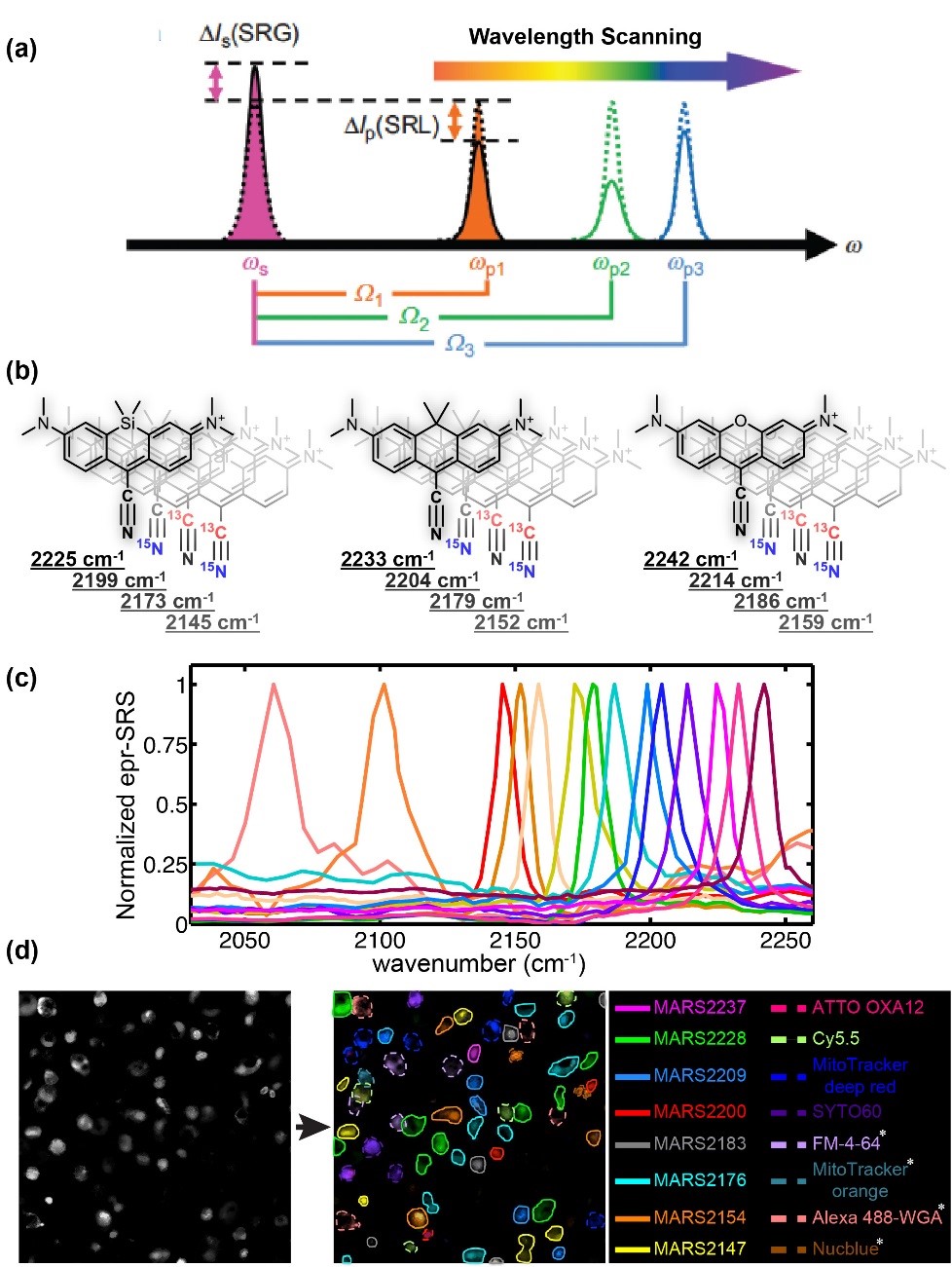
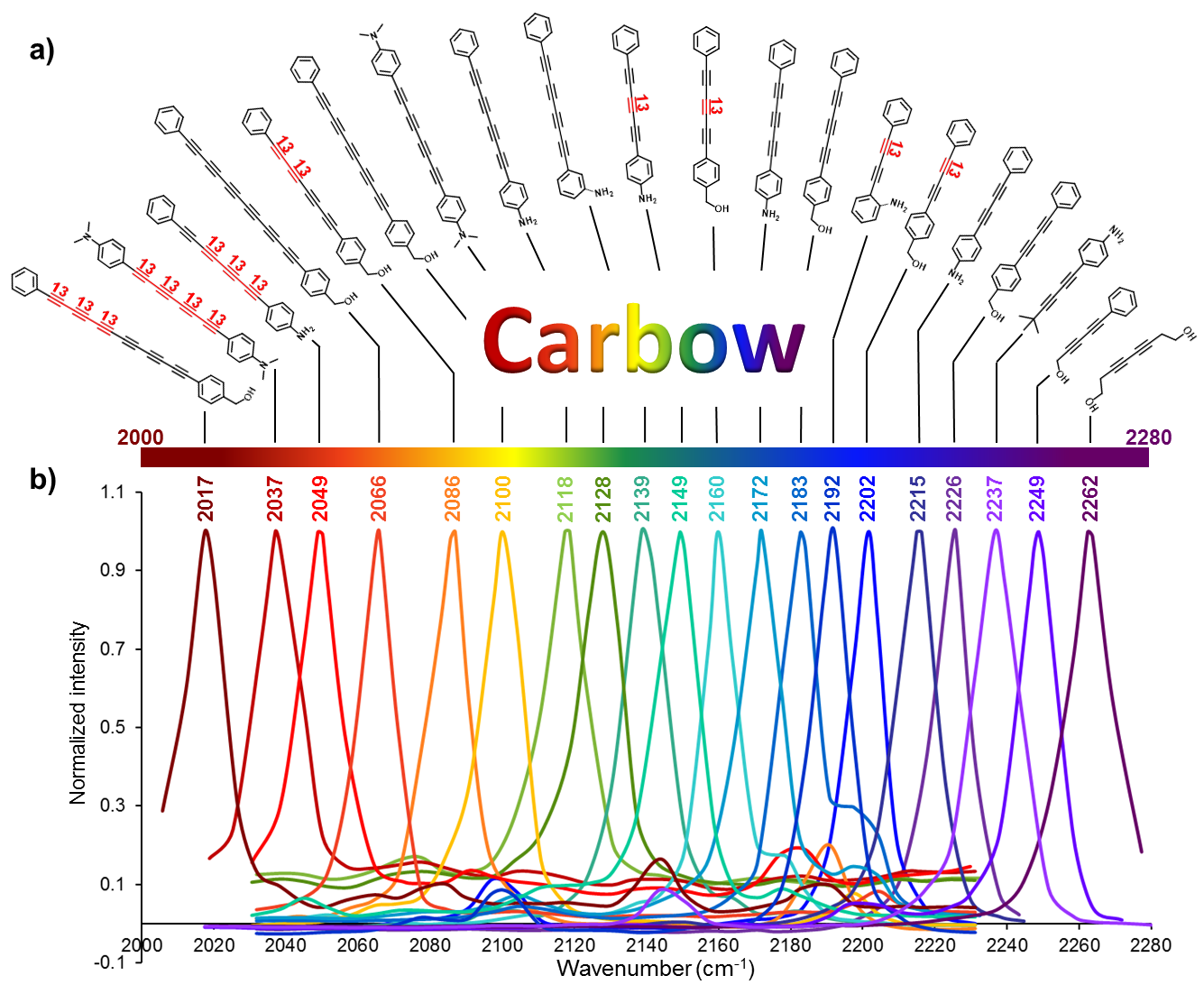
Engineered conjugated polyynes, or carbon-atom wires, provide a highly tunable palette of Raman probes. Varying chain length, isotopes, and end groups produced 20 spectrally distinct “Carbow” colors, greatly expanding the number of targets that can be resolved in a single sample.
- F. Hu, C. Zeng, R. Long, Y. Miao, L. Wei, Q. Xu and W. Min. “Super-multiplexed optical imaging and barcoding with engineered polyynes,” Nature Methods (2018).
- Z. Chen, D. Paley, L. Wei, A. Weisman, R. Friesner, C. Nuckolls* and W. Min*. “Multicolor live-cell chemical imaging by isotopically edited alkyne vibrational palette,” J. Am. Chem. Soc. 136, 8027 (2014).
Biological/Medical Applications
We apply SRS microscopy and bioorthogonal chemical imaging to reveal metabolism, molecular organization, and cellular identity across living cells, tissues, and organisms.
Imaging the Organelle Interactome in Living Cells
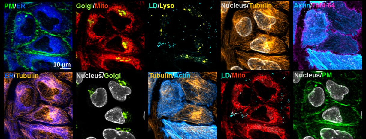
Cellular function emerges from continuous communication among membranes, organelles, and the cytoskeleton, yet conventional fluorescence imaging can observe only a few of these structures at once. To reveal this densely connected intracellular landscape, we combined five organelle-targeted Carbow Raman probes with five fluorescent reporters to achieve tandem ten-color imaging in living cells.
The resulting images simultaneously resolve the plasma membrane, endoplasmic reticulum, Golgi apparatus, mitochondria, lysosomes, lipid droplets, nucleus, tubulin, and actin. Because the Carbow probes occupy sharply separated Raman frequencies, these structures can be distinguished without computational unmixing or color compensation.
- F. Hu, C. Zeng, R. Long, Y. Miao, L. Wei, Q. Xu and W. Min. “Super-multiplexed optical imaging and barcoding with engineered polyynes,” Nature Methods (2018).
Lipid Metabolism
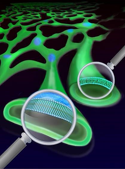
SRS tracking of deuterium-labeled lipids revealed that saturated-fat metabolites can form rigid, phase-separated domains in the endoplasmic reticulum. Unsaturated fatty acids can dissolve these domains, providing a physical mechanism that helps explain their protective effects in lipid-associated disorders.
- Y. Shen, Z. Zhao, L. Zhang, L. Shi, S. Shahriar, R. B. Chan, G. Paolo and W. Min. “Metabolic activity induces membrane phase separation in endoplasmic reticulum,” Proc. Natl. Acad. Sci. USA 114, 13394 (2017).
Protein Metabolism
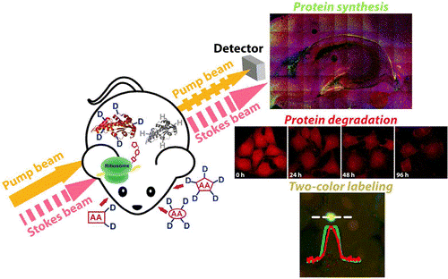
SRS imaging with deuterated amino acids enables noninvasive visualization of protein synthesis, degradation, trafficking, and pulse-chase dynamics in living systems. The method captures global protein turnover in cells, brain tissue, zebrafish, and mice without bulky fluorescent labels.
- L. Wei, Y. Shen, F. Xu, F. Hu, J. Harrington, K. Targoff and W. Min. “Imaging complex protein metabolism in live organisms by stimulated Raman scattering microscopy with isotope labeling,” ACS Chem. Biol. 10, 901 (2015).
- L. Wei, Y. Yu, Y. Shen, W. C. Wang* and W. Min*. “Vibrational imaging of newly synthesized proteins in live cells by stimulated Raman scattering microscopy,” Proc. Natl. Acad. Sci. USA 110, 11226 (2013).
- Y. Shen, F. Xu, L. Wei, F. Hu and W. Min. “Live-cell quantitative imaging of proteome degradation by stimulated Raman scattering,” Angew. Chem. Int. Ed. 53, 5596 (2014).
Glucose Metabolism
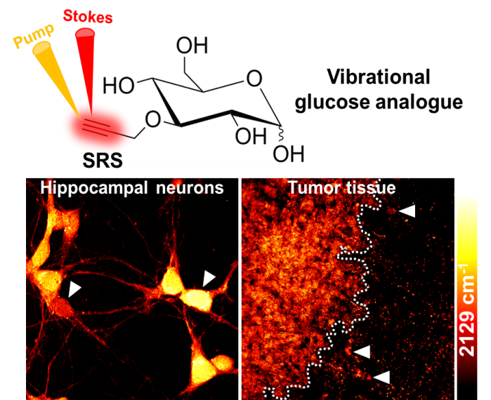
An alkyne-tagged glucose analogue and SRS microscopy map glucose uptake with optical and subcellular resolution. The approach distinguishes metabolic states and exposes cell-to-cell heterogeneity in tumors, neuronal cultures, and brain tissue without the spatial limitations of whole-body imaging methods.
- R. Long, L. Zhang, L. Shi, Y. Shen, F. Hu, C. Zeng and W. Min. “Two-color vibrational imaging of glucose metabolism by stimulated Raman scattering,” Chem. Commun. 54, 152 (2018).
- F. Hu, Z. Chen, L. Zhang, Y. Shen, L. Wei and W. Min. “Vibrational imaging of glucose uptake activity in live cells and tissues by stimulated Raman scattering,” Angew. Chem. Int. Ed. 54, 9821 (2015).
Cancer Metabolism
Bioorthogonal chemical imaging maps newly synthesized biomolecules in tumors after delivery of tagged metabolites in vivo. Its subcellular resolution reveals metabolic heterogeneity and helps dissect competition and cooperation among tumor, immune, and stromal cells.
- L. Zhang and W. Min. “Bioorthogonal chemical imaging of metabolic changes during epithelial–mesenchymal transition of cancer cells by stimulated Raman scattering microscopy,” J. Biomed. Opt. 22, 106010 (2017).
- F. Hu, Z. Chen, L. Zhang, Y. Shen, L. Wei and W. Min. “Vibrational imaging of glucose uptake activity in live cells and tissues by stimulated Raman scattering,” Angew. Chem. Int. Ed. 54, 9821 (2015).
Brain Metabolism
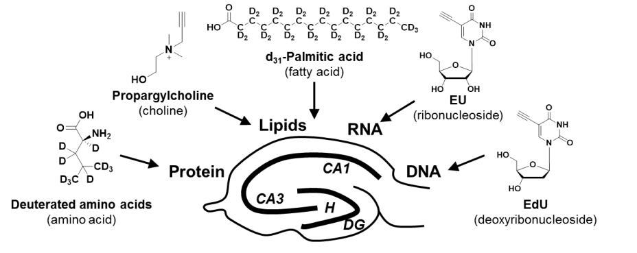
Integrated isotope and alkyne labeling with SRS microscopy visualizes DNA, RNA, protein, and lipid metabolism in living brain tissue. The platform resolves single-cell metabolic heterogeneity, neurogenesis, and localized responses to traumatic injury with minimal perturbation.
- F. Hu, M. R. Lamprecht, L. Wei, B. Morrison III and W. Min. “Bioorthogonal chemical imaging of metabolic activities in live mammalian hippocampal tissues with stimulated Raman scattering,” Scientific Reports 6, 39660 (2016).
- L. Wei, Y. Shen, F. Xu, F. Hu, J. Harrington, K. Targoff and W. Min. “Imaging complex protein metabolism in live organisms by stimulated Raman scattering microscopy with isotope labeling,” ACS Chem. Biol. 10, 901 (2015).